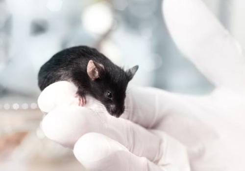
| Mice (wild-type) | SARS-CoV-2 (COVID-19) | SARS-CoV-1 (SARS) | MERS-CoV (MERS) | HCoV-229E (Seasonal) | HCoV-OC43 (Seasonal) | HCoV-NL63 (Seasonal) | HCoV-HKU1 (Seasonal) |
| Susceptibility | Not naturally susceptible: mouse ACE2 does not effectively bind SARS-CoV-2. | Partially permissive: mouse ACE2 does not efficiently bind SARS-CoV-1, but infection is achievable with high doses. Alternatively, a mouse-adapted SARS-CoV (MA15) was created to cause more robust infections in wild-type mice. | Not susceptible: murine DPP4 is non-functional for MERS. | Not naturally infected: wild-type mice resist HCoV-229E | Minimal: HCoV-OC43 can infect mice only if virus is adapted or mice are very young; standard models showed none to mild respiratory illness, with high dose inoculation producing greater disease. | Not susceptible: NL63 (uses ACE2) cannot infect wild-type mice (murine ACE2 not permissive, similar to SARS-CoVs). | Not susceptible: no small-animal model exists for HKU1 (it is difficult to culture and mouse tissues are not permissive). |
| Transmission | N/A (no infection) | In the MA15 mouse-adapted model, high viral titers in the lungs suggested high transmissibility, though this wasn’t explicitly studied. | N/A (no infection) | N/A (no infection) | Minimal (poor infection) | N/A (no infection) | N/A (no infection) |
| Shedding | N/A (no infection) | In the mouse-adapted virus models, pulmonary shedding is efficient, correlating with pulmonary titers. | N/A (no infection) | N/A (no infection) | Little to none | N/A (no infection) | N/A (no infection) |
| Symptoms and Pathology | N/A (no infection) | Young mice show virus replication in the lungs, but no overt signs of illness. Aged mice show mild symptoms. Adapted SARS-CoV strains cause severe lung pathology in mice without requiring transgenic modification. | N/A (no infection) | N/A (no infection) | Possibly mild or neurotrophic effects in infant mice, but not significant respiratory disease. | N/A (no infection) | N/A (no infection) |
| Ethics and Logistics | N/A (no infection) | Wild-type mice are cheap and available, and infection with adapted virus provides a useful model. BSL-3 is required. | N/A (no infection) | N/A (no infection) | Wild type mice are cheap and available, but of little use for HCoV-OC43. | Wild type mice are cheap and available, but of little use for HCoV-NL63. | Wild type mice are cheap and available, but of little use for HCoV-HKU1. |
Mice (wild-type): References
Dangi, Tanushree, Shiyi Li, and Pablo Penaloza-MacMaster. 2025. “Development of a Cross-Protective Common Cold Coronavirus Vaccine.” Journal of Virology, October 22, e0152625. https://doi.org/10.1128/jvi.01526-25
Dinnon, Kenneth H., Sarah R. Leist, Alexandra Schäfer, Caitlin E. Edwards, David R. Martinez, Stephanie A. Montgomery, Ande West, et al. 2020. “A Mouse-Adapted Model of SARS-CoV-2 to Test COVID-19 Countermeasures.” Nature 586 (7830): 560–66. https://doi.org/10.1038/s41586-020-2708-8.
Liu, Donglan, Chunke Chen, Dingbin Chen, Airu Zhu, Fang Li, Zhen Zhuang, Chris Ka Pun Mok, et al. 2023. “Mouse Models Susceptible to HCoV-229E and HCoV-NL63 and Cross Protection from Challenge with SARS-CoV-2.” Proceedings of the National Academy of Sciences of the United States of America 120 (4): e2202820120. https://doi.org/10.1073/pnas.2202820120.
Roberts, Anjeanette, Damon Deming, Christopher D. Paddock, Aaron Cheng, Boyd Yount, Leatrice Vogel, Brian D. Herman, et al. 2007. “A Mouse-Adapted SARS-Coronavirus Causes Disease and Mortality in BALB/c Mice.” PLOS Pathogens 3 (1): e5. https://doi.org/10.1371/journal.ppat.0030005.
Shou, Shuyu, Menghui Liu, Yang Yang, Ning Kang, Yingying Song, Dan Tan, Nannan Liu, Feifei Wang, Jing Liu, and Youhua Xie. 2021. “Animal Models for COVID-19: Hamsters, Mouse, Ferret, Mink, Tree Shrew, and Non-Human Primates.” Frontiers in Microbiology 12 (August):626553. https://doi.org/10.3389/fmicb.2021.626553.
Xie, Peifang, Yue Fang, Zulqarnain Baloch, Huanhuan Yu, Zeyuan Zhao, Rongqiao Li, Tongtong Zhang, et al. 2022. “A Mouse-Adapted Model of HCoV-OC43 and Its Usage to the Evaluation of Antiviral Drugs.” Frontiers in Microbiology 13:845269. https://doi.org/10.3389/fmicb.2022.845269.
| Mice (engineered) | SARS-CoV-2 (COVID-19) | SARS-CoV-1 (SARS) | MERS-CoV (MERS) | HCoV-229E (Seasonal) | HCoV-OC43 (Seasonal) | HCoV-NL63 (Seasonal) | HCoV-HKU1 (Seasonal) |
| Susceptibility | Transgenic mice expressing human ACE2 are susceptible to SARS-CoV-2, supporting infection of the airways. Mouse-adapted SARS-CoV-2 has also been developed to improve infection. | hACE2-transgenic mice permit SARS-CoV infection in airway cells. | Transgenic mice expressing human DPP4 are fully susceptible to MERS-CoV. Also, mice transiently “humanized” with an adenovirus carrying hDPP4 can be infected. Serial passage of MERS-CoV through human DPP4 knock-in or CRISPR-edited mice have yielded adapted strains (MERSMA 6.1.2, MERS-15/icMERSma1, maM35c4) that can be highly virulent in these mouse lines. | Yes (with modifications). Mice can be made susceptible to 229E by delivering human APN (the 229E receptor) via adenovirus, usually in IFN receptor–knockout backgrounds. | Researchers have serially passaged HCoV-OC43 in mice to create a mouse-adapted OC43 strain. | Similar to 229E, an adenovirus delivering human ACE2 renders mice (particularly IFNAR–/–) susceptible to NL63. | No transgenic or adapted mouse model has been successfully developed for HKU1 to date. (HKU1’s cellular receptor and growth requirements make modeling difficult.) |
| Transmission | In K18-hACE2 or similar models, virus can spread to naïve mice via close contact and respiratory droplets. Aerosol-only exposure is less efficient without prolonged high-dose exposure. | Engineered mice are mainly used to study pathology; direct transmission studies are rare. (High ACE2 expression in some models caused fatal encephalitis, complicating transmission experiments.) | MERS in hDPP4 mice causes severe disease, and mice typically succumb before transmission can be assessed. (Mouse-to-mouse transmission has not been a focus due to acute lethality). | Poorly studied: these models are primarily used to study immunity and interventions, not natural transmission. (Virus is cleared ~7 days post-infection). | Not typically measured: studies focus on disease and treatment. (Adapted OC43 causes rapid disease in newborn mice, so any transmission would be limited by quick host death.) | Used mainly for immune response studies, not for studying transmission dynamics. (Virus is cleared within ~7 days) | N/A (no mouse model) |
| Shedding | In hACE2 mice, virus replication is primarily in lungs/nasal tract; shedding of infectious virus occurs for a few days post-infection (detailed shedding kinetics in mice are less reported than in hamsters). | In hACE2 mice, virus replicates in lungs; shedding of SARS-CoV has been confirmed in respiratory tissues but not extensively quantified in literature (focus was on disease outcome). | High virus titers in lungs; infected hDPP4 mice shed virus (e.g. detectable in respiratory secretions), but die quickly, limiting shedding duration. | Virus replicates in lungs; infectious virus is present for several days and then clears. Shedding likely occurs via respiratory secretions during that period, though specific droplet transmission in mice has not been reported. | The adapted OC43 replicates to high titers in lungs. Infected mice likely shed virus in respiratory secretions until death (≤5 days post-infection) | Virus replicates in lungs and likely nasal tissues for less than a week in these mice. Active shedding would be limited to the first week post-infection. | N/A (no mouse model) |
| Symptoms and Pathology | Mild to moderate pneumonia and weight loss occur. Some hACE2 models (e.g., overexpressing ACE2) also develop brain infection leading to neurological signs. | hACE2 mice develop pneumonia and in some cases severe disease (even death) due to viral spread to CNS. | Severe pneumonia and often fatal outcomes occur in hDPP4-transgenic mice, especially when using adapted strains, mirroring serious human MERS. | In immunodeficient (STAT1–/– or IFNAR–/–) mice with hAPN, 229E infection causes pneumonia with inflammatory infiltrates, but mice recover by 1 week. | The mouse-adapted OC43 causes significant weight loss and death in neonatal mice, with severe viral pneumonia and encephalitis observed. Cytokine storms and lung pathology mimic severe outcomes in vulnerable humans. | NL63-infected hACE2-transduced mice develop pneumonia with inflammatory cell infiltration, though disease is usually moderate and resolves with viral clearance by day 7. | N/A (no mouse model) |
| Ethics and Logistics | Transgenic lines (or adenovirus-hACE2 transduction) require specialized breeding, but mice remain a convenient, low-cost model for SARS-CoV-2 with human-like receptors. | These models enabled SARS research but involve genetic modification or adapted high-virulence virus; require high-level containment and careful monitoring for neuro symptoms. BSL-3 is required. | These models are crucial for MERS studies, but transgenic breeding or viral transduction is needed. Infected mice must be handled in high-containment due to the severe disease and high viral loads. BSL-3 is required. | Requires adenovirus transduction and immunodeficient mice. This is a specialized model allowing vaccine/antiviral testing and showing cross-protection. | Useful for antiviral drug testing, but lethality (unlike in humans) limits its utility for other study types. | Requires adenoviral gene delivery plus immunodeficient mice. Useful for testing NL63-targeted therapies/vaccines and studying cross-reactive immunity. | N/A (no mouse model) |
Mice (engineered): References
Agrawal, Anurodh Shankar, Tania Garron, Xinrong Tao, Bi-Hung Peng, Maki Wakamiya, Teh-Sheng Chan, Robert B. Couch, and Chien-Te K. Tseng. 2015. “Generation of a Transgenic Mouse Model of Middle East Respiratory Syndrome Coronavirus Infection and Disease.” Journal of Virology 89 (7): 3659–70. https://doi.org/10.1128/JVI.03427-14.
Bi, Zhenfei, Weiqi Hong, Jingyun Yang, Shuaiyao Lu, and Xiaozhong Peng. 2021. “Animal Models for SARS‐CoV‐2 Infection and Pathology.” MedComm 2 (4): 548–68. https://doi.org/10.1002/mco2.98.
Caldera-Crespo, Luis A., Michael J. Paidas, Sabita Roy, Carl I. Schulman, Norma Sue Kenyon, Sylvia Daunert, and Arumugam R. Jayakumar. 2022. “Experimental Models of COVID-19.” Frontiers in Cellular and Infection Microbiology 11. https://www.frontiersin.org/article/10.3389/fcimb.2021.792584.
Casel, Mark Anthony B., Rare G. Rollon, and Young Ki Choi. 2021. “Experimental Animal Models of Coronavirus Infections: Strengths and Limitations.” Immune Network 21 (2): e12. https://doi.org/10.4110/in.2021.21.e12.
Cockrell, Adam S., Boyd L. Yount, Trevor Scobey, et al. 2017. “A Mouse Model for MERS Coronavirus-Induced Acute Respiratory Distress Syndrome.” Nature Microbiology 2 (2): 16226. https://doi.org/10.1038/nmicrobiol.2016.226.
Douglas, Madeline G., Jacob F. Kocher, Trevor Scobey, Ralph S. Baric, and Adam S. Cockrell. 2018. “Adaptive Evolution Influences the Infectious Dose of MERS-CoV Necessary to Achieve Severe Respiratory Disease.” Virology 517 (April): 98–107. https://doi.org/10.1016/j.virol.2017.12.006.
Lassnig, Caroline, Carlos M. Sanchez, Monika Egerbacher, Ingrid Walter, Susanne Majer, Thomas Kolbe, Pilar Pallares, Luis Enjuanes, and Mathias Müller. 2005. “Development of a Transgenic Mouse Model Susceptible to Human Coronavirus 229E.” Proceedings of the National Academy of Sciences of the United States of America 102 (23): 8275–80. https://doi.org/10.1073/pnas.0408589102.
Lee, Chung-Young, and Anice C Lowen. 2021. “Animal Models for SARS-CoV-2.” Current Opinion in Virology 48 (June):73–81. https://doi.org/10.1016/j.coviro.2021.03.009.
Li, Kun, Christine L. Wohlford-Lenane, Rudragouda Channappanavar, et al. 2017. “Mouse-Adapted MERS Coronavirus Causes Lethal Lung Disease in Human DPP4 Knockin Mice.” Proceedings of the National Academy of Sciences of the United States of America 114 (15): E3119–28. https://doi.org/10.1073/pnas.1619109114.
Liu, Donglan, Chunke Chen, Dingbin Chen, Airu Zhu, Fang Li, Zhen Zhuang, Chris Ka Pun Mok, et al. 2023. “Mouse Models Susceptible to HCoV-229E and HCoV-NL63 and Cross Protection from Challenge with SARS-CoV-2.” Proceedings of the National Academy of Sciences of the United States of America 120 (4): e2202820120. https://doi.org/10.1073/pnas.2202820120.
McCray, Paul B., Lecia Pewe, Christine Wohlford-Lenane, Melissa Hickey, Lori Manzel, Lei Shi, Jason Netland, et al. 2007. “Lethal Infection of K18-hACE2 Mice Infected with Severe Acute Respiratory Syndrome Coronavirus.” Journal of Virology 81 (2): 813–21. https://doi.org/10.1128/JVI.02012-06.
Muñoz-Fontela, César, Lina Widerspick, Randy A. Albrecht, Martin Beer, Miles W. Carroll, Emmie de Wit, Michael S. Diamond, et al. 2022. “Advances and Gaps in SARS-CoV-2 Infection Models.” PLOS Pathogens 18 (1): e1010161. https://doi.org/10.1371/journal.ppat.1010161.
Sefik, Esen, Ben Israelow, Jun Zhao, Rihao Qu, Eric Song, Haris Mirza, Eleanna Kaffe, et al. 2021. “A Humanized Mouse Model of Chronic COVID-19 to Evaluate Disease Mechanisms and Treatment Options.” Research Square, March, rs.3.rs-279341. https://doi.org/10.21203/rs.3.rs-279341/v1.
Shou, Shuyu, Menghui Liu, Yang Yang, Ning Kang, Yingying Song, Dan Tan, Nannan Liu, Feifei Wang, Jing Liu, and Youhua Xie. 2021. “Animal Models for COVID-19: Hamsters, Mouse, Ferret, Mink, Tree Shrew, and Non-Human Primates.” Frontiers in Microbiology 12 (August):626553. https://doi.org/10.3389/fmicb.2021.626553.
Zhang, Liang, Shuaiyin Chen, Weiguo Zhang, Haiyan Yang, Yuefei Jin, and Guangcai Duan. 2021. “An Update on Animal Models for Severe Acute Respiratory Syndrome Coronavirus 2 Infection and Countermeasure Development.” Frontiers in Microbiology 12 (November):770935. https://doi.org/10.3389/fmicb.2021.770935.
Zhao, Jincun, Kun Li, Christine Wohlford-Lenane, Sudhakar S. Agnihothram, Craig Fett, Jingxian Zhao, Michael J. Gale, et al. 2014. “Rapid Generation of a Mouse Model for Middle East Respiratory Syndrome.” Proceedings of the National Academy of Sciences of the United States of America 111 (13): 4970–75. https://doi.org/10.1073/pnas.1323279111.
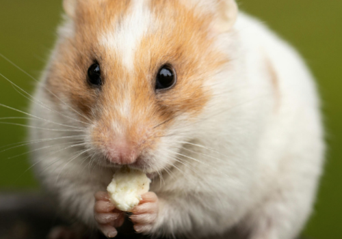
| Hamsters (Syrian golden) | SARS-CoV-2 (COVID-19) | SARS-CoV-1 (SARS) | MERS-CoV (MERS) | HCoV-229E (Seasonal) | HCoV-OC43 (Seasonal) | HCoV-NL63 (Seasonal) | HCoV-HKU1 (Seasonal) |
| Susceptibility | Golden Syrian hamsters are highly susceptible to SARS-CoV-2, with the virus efficiently infecting the nasal mucosa, bronchial epithelium, and lungs. | Hamsters support SARS-CoV-1 replication similarly to SARS-CoV-2 (permissive ACE2 receptor). | Not susceptible: Hamster DPP4 is non-functional for MERS. | Hamsters are not commonly used for 229E studies (hamster APN may not support 229E; this has not been a focus). | There is little to no literature on OC43 infection in hamsters; hamsters are not a standard OC43 model. | No notable reports of NL63 infection in hamsters; not a standard model. | N/A (no established hamster model) |
| Transmission | Infected hamsters readily transmit SARS-CoV-2 to naïve cage mates through direct contact and via airborne droplets/aerosols.10 In experiments, all contact hamsters and even some separated by airflow become infected. Fomite transmission is less efficient. Some Omicron (BA.1 and BA.2) strains may be less transmissible in hamster models. | While direct transmission experiments are not extensively reported, hamsters shed SARS-CoV (high lung titers) which suggests they could transmit if cohoused. (By analogy to SARS-2, hamsters are likely capable of droplet spread.) | N/A (no infection) | Not studied | Not studied | Not studied | N/A (no hamster model) |
| Shedding | Hamsters shed high levels of virus from the upper airway. Viral RNA is detectable in nasal washes for ~14 days, though infectious virus is shed mostly in the first week. | Peak lung virus occurs at day 2 and is rapidly cleared by day 7. Shedding from the respiratory tract would be brief. No weight loss or clinical illness was seen with the Urbani strain, but a more virulent SARS strain (Frk-1) caused lethality in hamsters. | N/A (no infection) | Not studied | Not studied | Not studied | N/A (no hamster model) |
| Symptoms and Pathology | Infected hamsters experience weight loss and recover within 2 weeks. They develop lung lesions (pneumonia with lung consolidation) similar to mild human COVID-19. Hamsters efficiently mount neutralizing antibody responses and clear the virus. Some Omicron strains (BA.1 and BA.2) may induce more limited disease than lineages (particularly Delta and Gamma). | Urbani strain caused subclinical infection (no obvious disease) in hamsters despite robust viral replication. Certain strains can cause severe disease (hamsters infected with Frk-1 experienced lethal outcomes). | N/A (no infection) | Not studied | Not studied | Not studied | N/A (no hamster model) |
| Ethics and Logistics | Hamsters are small and easy to handle; infection causes self-limited illness (ethically sound). They have been a workhorse model for COVID-19 transmission and pathogenesis studies. | Hamsters were used in some SARS studies; they are inexpensive and easy, and pose minimal ethical concerns due to typically mild disease (depending on strain). However, the mild illness in hamsters with standard SARS-CoV strains limits their use for pathogenesis studies. BSL-3 is required. | N/A (no infection) | Hamsters are generally not used for 229E studies. | Hamsters are generally not used for OC43 studies. | Hamsters are generally not used for NL63 studies. | N/A (no hamster model) |
Hamsters (Syrian golden): References
Bi, Zhenfei, Weiqi Hong, Jingyun Yang, Shuaiyao Lu, and Xiaozhong Peng. 2021. “Animal Models for SARS‐CoV‐2 Infection and Pathology.” MedComm 2 (4): 548–68. https://doi.org/10.1002/mco2.98.
Braxton, Alicia M, Patrick S Creisher, Camilo A Ruiz-Bedoya, Katie R Mulka, Santosh Dhakal, Alvaro A Ordonez, Sarah E Beck, Sanjay K Jain, and Jason S Villano. “Hamsters as a Model of Severe Acute Respiratory Syndrome Coronavirus-2.” Comparative Medicine 71, no. 5 (October 2021): 1–13. https://doi.org/10.30802/AALAS-CM-21-000036.
Caldera-Crespo, Luis A., Michael J. Paidas, Sabita Roy, Carl I. Schulman, Norma Sue Kenyon, Sylvia Daunert, and Arumugam R. Jayakumar. 2022. “Experimental Models of COVID-19.” Frontiers in Cellular and Infection Microbiology 11. https://www.frontiersin.org/article/10.3389/fcimb.2021.792584.
Casel, Mark Anthony B., Rare G. Rollon, and Young Ki Choi. 2021. “Experimental Animal Models of Coronavirus Infections: Strengths and Limitations.” Immune Network 21 (2): e12. https://doi.org/10.4110/in.2021.21.e12.
Choudhary, Shambhunath, Isis Kanevsky, Soner Yildiz, Rani S. Sellers, Kena A. Swanson, Tania Franks, Raveen Rathnasinghe, et al. “Modeling SARS-CoV-2: Comparative Pathology in Rhesus Macaque and Golden Syrian Hamster Models.” Toxicologic Pathology, February 5, 2022, 01926233211072767. https://doi.org/10.1177/01926233211072767.
Frere, Justin J., Randal A. Serafini, Kerri D. Pryce, Marianna Zazhytska, Kohei Oishi, Ilona Golynker, Maryline Panis, et al. “SARS-CoV-2 Infection in Hamsters and Humans Results in Lasting and Unique Systemic Perturbations Post Recovery.” Science Translational Medicine 0, no. 0 (June 7, 2022): eabq3059. https://doi.org/10.1126/scitranslmed.abq3059.
Halfmann, Peter J., Shun Iida, Kiyoko Iwatsuki-Horimoto, Tadashi Maemura, Maki Kiso, Suzanne M. Scheaffer, Tamarand L. Darling, et al. “SARS-CoV-2 Omicron Virus Causes Attenuated Disease in Mice and Hamsters.” Nature, January 21, 2022. https://doi.org/10.1038/s41586-022-04441-6.
Imai, Masaki, Kiyoko Iwatsuki-Horimoto, Masato Hatta, Samantha Loeber, Peter J. Halfmann, Noriko Nakajima, Tokiko Watanabe, et al. “Syrian Hamsters as a Small Animal Model for SARS-CoV-2 Infection and Countermeasure Development.” Proceedings of the National Academy of Sciences of the United States of America 117, no. 28 (July 14, 2020): 16587–95. https://doi.org/10.1073/pnas.2009799117.
Lee, Chung-Young, and Anice C Lowen. 2021. “Animal Models for SARS-CoV-2.” Current Opinion in Virology 48 (June):73–81. https://doi.org/10.1016/j.coviro.2021.03.009.
Muñoz-Fontela, César, Lina Widerspick, Randy A. Albrecht, Martin Beer, Miles W. Carroll, Emmie de Wit, Michael S. Diamond, et al. 2022. “Advances and Gaps in SARS-CoV-2 Infection Models.” PLOS Pathogens 18 (1): e1010161. https://doi.org/10.1371/journal.ppat.1010161.
Shou, Shuyu, Menghui Liu, Yang Yang, Ning Kang, Yingying Song, Dan Tan, Nannan Liu, Feifei Wang, Jing Liu, and Youhua Xie. “Animal Models for COVID-19: Hamsters, Mouse, Ferret, Mink, Tree Shrew, and Non-Human Primates.” Frontiers in Microbiology 12 (August 31, 2021): 626553. https://doi.org/10.3389/fmicb.2021.626553.
Sia, Sin Fun, Li-Meng Yan, Alex WH Chin, Kevin Fung, Ka-Tim Choy, Alvina YL Wong, Prathanporn Kaewpreedee, et al. “Pathogenesis and Transmission of SARS-CoV-2 in Golden Syrian Hamsters.” Nature 583, no. 7818 (July 2020): 834–38. https://doi.org/10.1038/s41586-020-2342-5.
Wuertz, Kathryn McGuckin, Erica K. Barkei, Wei-Hung Chen, Elizabeth J. Martinez, Ines Lakhal-Naouar, Linda L. Jagodzinski, Dominic Paquin-Proulx, et al. “A SARS-CoV-2 Spike Ferritin Nanoparticle Vaccine Protects Hamsters against Alpha and Beta Virus Variant Challenge.” NPJ Vaccines 6 (October 28, 2021): 129. https://doi.org/10.1038/s41541-021-00392-7.
Zhang, Liang, Shuaiyin Chen, Weiguo Zhang, Haiyan Yang, Yuefei Jin, and Guangcai Duan. 2021. “An Update on Animal Models for Severe Acute Respiratory Syndrome Coronavirus 2 Infection and Countermeasure Development.” Frontiers in Microbiology 12 (November):770935. https://doi.org/10.3389/fmicb.2021.770935.
Banete, Andra, Bryan D. Griffin, Juan C. Corredor, Emily Chien, Lily Yip, Tarini N. A. Gunawardena, Kuganya Nirmalarajah, et al. 2025. “Pathogenesis and Transmission of SARS-CoV-2 D614G, Alpha, Gamma, Delta, and Omicron Variants in Golden Hamsters.” Npj Viruses 3 (1): 15. https://doi.org/10.1038/s44298-025-00092-2.
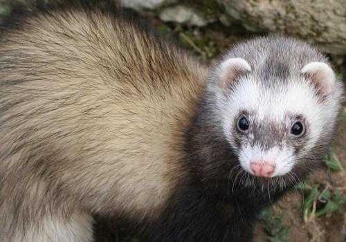
| Ferrets | SARS-CoV-2 (COVID-19) | SARS-CoV-1 (SARS) | MERS-CoV (MERS) | HCoV-229E (Seasonal) | HCoV-OC43 (Seasonal) | HCoV-NL63 (Seasonal) | HCoV-HKU1 (Seasonal) |
| Susceptibility | Ferrets are permissive to SARS-CoV-2 infection, though they typically exhibit mild clinical illness. The virus replicates in the upper respiratory tract. | Ferrets are susceptible to SARS-CoV-1; experiments have shown ferrets become infected, and often exhibit severe illness. | Ferrets are not susceptible to MERS-CoV. Experimental inoculation with high-dose MERS-CoV via intranasal / tracheal routes resulted in only transient viral RNA detection in ferret swabs, with no infectious virus recovered. | No known model. Ferrets are not typically infected by 229E (a human cold virus). There is no literature on successful ferret infection with HCoV-229E. | Ferrets are not known to contract OC43. (Ferrets have their own coronaviruses, but human OC43 has not been reported to infect ferrets.) | NL63 has not been demonstrated to infect ferrets. | HKU1 has not been demonstrated to infect ferrets. |
| Transmission | Ferrets readily transmit SARS-CoV-2 via respiratory droplets and possibly aerosols. In experiments, infected ferrets transmitted virus to direct contacts and even to ferrets in separate cages over >1 meter distance. This demonstrates airborne droplet spread in this model. | Infected ferrets shed SARS-CoV and can infect naive ferrets by direct contact. In one study, uninfected ferrets housed with infected ones became PCR-positive within 2 days. Ferrets have also transmitted SARS-CoV via the air in experimental setups (all 4 indirect-contact ferrets became infected in one test). | N/A (no infection) | N/A (no ferret model) | Not studied | Not studied | Not studied |
| Shedding | Ferrets shed infectious virus from the nasopharynx for ~7-10 days. SARS-CoV-2 has been detected in ferret nasal washes within 2 days of infection. Ferret-to-ferret transmission has been noted to occur within 2 days of exposure, indicating early and robust shedding. | Similar to SARS-CoV-2, ferrets shed SARS-CoV-1 from the throat/nose starting ~2 days post-inoculation and continuing for ~14 days. High viral titers were found in ferret lungs and upper airways. | None (only trace RNA for ~2 days, no live virus) | N/A (no ferret model) | Not studied | Not studied | Not studied |
| Symptoms and Pathology | Infected ferrets usually develop mild symptoms. Disease tends to be self-limited without severe lung pathology. Ferrets rarely lose much weight or die from SARS-2; pathology is often limited to mild rhinitis or bronchitis. | Infected ferrets generally show mild disease, but some exhibit more advanced symptoms (e.g., lethargy, conjunctivitis, lung lesions, weight loss, rarely death). Very little signs of classical SARS pneumonia. | No illness has been seen, since MERS-CoV does not replicate in ferret cells. | N/A (no ferret model) | Not studied | Not studied | Not studied |
| Ethics and Logistics | Ferrets are a classic respiratory virus model (similar airway physiology to humans). They require more space and specialized care than rodents. Ferrets generally tolerate infection well (ethical impact is low), making them ideal for transmission experiments. | Ferrets were validated as a SARS model and used to test vaccines/therapeutics. Disease severity can be moderate, so monitoring and humane endpoints are needed. BSL-3 is required. | Ferrets are a poor model for MERS, and BSL-3 is required. | N/A (no ferret model) | Not used | Not used | Not used |
Ferrets: References
Bi, Zhenfei, Weiqi Hong, Jingyun Yang, Shuaiyao Lu, and Xiaozhong Peng. 2021. “Animal Models for SARS‐CoV‐2 Infection and Pathology.” MedComm 2 (4): 548–68. https://doi.org/10.1002/mco2.98.
Caldera-Crespo, Luis A., Michael J. Paidas, Sabita Roy, Carl I. Schulman, Norma Sue Kenyon, Sylvia Daunert, and Arumugam R. Jayakumar. 2022. “Experimental Models of COVID-19.” Frontiers in Cellular and Infection Microbiology 11. https://www.frontiersin.org/article/10.3389/fcimb.2021.792584.
Casel, Mark Anthony B., Rare G. Rollon, and Young Ki Choi. 2021. “Experimental Animal Models of Coronavirus Infections: Strengths and Limitations.” Immune Network 21 (2): e12. https://doi.org/10.4110/in.2021.21.e12.
Chu, Yong-Kyu, Georgia D. Ali, Fuli Jia, Qianjun Li, David Kelvin, Ronald C. Couch, Kevin S. Harrod, et al. 2008. “The SARS-CoV Ferret Model in an Infection–Challenge Study.” Virology 374 (1): 151–63. https://doi.org/10.1016/j.virol.2007.12.032.
Ciurkiewicz, Malgorzata, Federico Armando, Tom Schreiner, Nicole de Buhr, Veronika Pilchová, Vanessa Krupp-Buzimikic, Gülşah Gabriel, et al. 2022. “Ferrets Are Valuable Models for SARS-CoV-2 Research.” Veterinary Pathology, January, 03009858211071012. https://doi.org/10.1177/03009858211071012.
Kutter, Jasmin S., Dennis de Meulder, Theo M. Bestebroer, Pascal Lexmond, Ard Mulders, Mathilde Richard, Ron A. M. Fouchier, and Sander Herfst. 2021. “SARS-CoV and SARS-CoV-2 Are Transmitted through the Air between Ferrets over More than One Meter Distance.” Nature Communications 12 (1): 1653. https://doi.org/10.1038/s41467-021-21918-6.
Lee, Chung-Young, and Anice C Lowen. 2021. “Animal Models for SARS-CoV-2.” Current Opinion in Virology 48 (June):73–81. https://doi.org/10.1016/j.coviro.2021.03.009.
Muñoz-Fontela, César, Lina Widerspick, Randy A. Albrecht, Martin Beer, Miles W. Carroll, Emmie de Wit, Michael S. Diamond, et al. 2022. “Advances and Gaps in SARS-CoV-2 Infection Models.” PLOS Pathogens 18 (1): e1010161. https://doi.org/10.1371/journal.ppat.1010161.
Shou, Shuyu, Menghui Liu, Yang Yang, Ning Kang, Yingying Song, Dan Tan, Nannan Liu, Feifei Wang, Jing Liu, and Youhua Xie. 2021. “Animal Models for COVID-19: Hamsters, Mouse, Ferret, Mink, Tree Shrew, and Non-Human Primates.” Frontiers in Microbiology 12 (August):626553. https://doi.org/10.3389/fmicb.2021.626553.
Witt, Alexandra N, Rachel D Green, and Andrew N Winterborn. 2021. “A Meta-Analysis of Rhesus Macaques (Macaca Mulatta), Cynomolgus Macaques (Macaca Fascicularis), African Green Monkeys (Chlorocebus Aethiops), and Ferrets (Mustela Putorius Furo) as Large Animal Models for COVID-19.” Comparative Medicine 71 (5): 1–9. https://doi.org/10.30802/AALAS-CM-21-000032.
Zhang, Liang, Shuaiyin Chen, Weiguo Zhang, Haiyan Yang, Yuefei Jin, and Guangcai Duan. 2021. “An Update on Animal Models for Severe Acute Respiratory Syndrome Coronavirus 2 Infection and Countermeasure Development.” Frontiers in Microbiology 12 (November):770935. https://doi.org/10.3389/fmicb.2021.770935.
| Non-human primates | SARS-CoV-2 (COVID-19) | SARS-CoV-1 (SARS) | MERS-CoV (MERS) | HCoV-229E (Seasonal) | HCoV-OC43 (Seasonal) | HCoV-NL63 (Seasonal) | HCoV-HKU1 (Seasonal) |
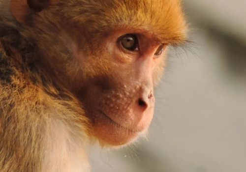
| Susceptibility | Multiple NHP species are susceptible to SARS-CoV-2. Rhesus macaques and cynomolgus macaques develop infection with viral replication in the respiratory tract. Disease severity is typically mild-to-moderate (e.g. rhesus macaques show moderate pneumonia and clinical signs, cynomolgus have milder symptoms). African green monkeys may exhibit more pronounced disease in some studies. | Rhesus macaques were infected with SARS-CoV-1 during the SARS outbreak investigations. They developed an illness with fever and lung involvement. NHP infection was productive, and viral replication in lungs was confirmed. | NHPs are susceptible to MERS-CoV, with species differences: Rhesus macaques develop a mild-to-moderate respiratory illness (rhinitis, cough, mild pneumonia), whereas common marmosets (small New World primates) develop severe disease more akin to lethal human cases. Marmosets often progress to severe pneumonia and even death, while macaques recover. | A novel NHP model for HCoV-229E in pigtailed macaques was developed, which mimics the mild upper-respiratory disease seen in humans. The macaques showed cold-like symptoms and were used to examine immunity and cross-protection. | Some NHPs are susceptible, but their use is very limited due to lack of ethical and budgetary justification. | Some NHPs are susceptible, but their use is very limited due to lack of ethical and budgetary justification. | Some NHPs are susceptible, but their use is very limited due to lack of ethical and budgetary justification. |
| Transmission | NHPs are generally not used for transmission experiments due to cost and ethical constraints. In research settings, infected NHPs are usually housed separately; thus, in-cage transmission is not usually studied. (The focus is on pathology and therapeutic testing rather than transmission). | Not typically studied in NHPs. (During SARS, transmission dynamics were studied in small animals; NHP experiments were focused on pathology/vaccine). Cohousing infected and uninfected macaques is rarely done due to risk, therefore, we have no clear data on NHP-to-NHP droplet spread for SARS, though it’s plausible if they were in proximity. | Not studied in NHPs (each is usually infected intentionally). No reported NHP-to-NHP transmission experiments for MERS. | Not extensively studied | Not extensively studied | Not extensively studied | Not extensively studied |
| Shedding | Infected macaques shed SARS-CoV-2 from the nose and throat (swabs detect virus RNA). Shedding peaks a few days post-infection and then declines as the monkeys clear the virus, usually within 1-2 weeks. NHPs can also shed virus in bronchial fluids and occasionally feces (reflecting systemic spread). | Infected macaques shed SARS-CoV-1 in nasal and tracheal secretions. Shedding likely peaks around day 2-4 post infection (as fever spikes) and then decreases as the immune response controls the virus. | Infected rhesus macaques shed MERS-CoV from the upper airways for a short period (virus in nasal swabs), clearing the infection relatively quickly. Marmosets with severe disease likely shed higher titers and for longer, but due to ethical endpoints (euthanasia when moribund), prolonged shedding data are limited. | In the 229E macaque model, virus was shed in nasal secretions for a short period (a few days) as the animal experienced a self-limited “cold.” Viral loads were much lower than those observed with SARS or MERS in NHPs. | Not extensively studied | Not extensively studied | Not extensively studied |
| Symptoms and Pathology | Macaques develop fever, reduced appetite, and radiographic lung infiltrates. Histologically, they get viral pneumonia (alveolar edema, inflammatory infiltrates) akin to mild human COVID-19. They generally do not progress to ARDS or death. All recover and develop antibodies. | Macaques develop pneumonia similar to human SARS, including diffuse alveolar damage in lungs. Clinical signs are moderate; most macaques did not die but showed significant lung lesions on necropsy. The pathology in macaques helps confirm SARS-CoV-1 as the cause of SARS in humans. | Rhesus macaque MERS is characterized by transient fever and lung inflammation; a closer model of mild human MERS. In contrast, marmosets show weight loss, severe lung lesions, extensive viral pneumonia, and can experience organ failure, mirroring the ~35–40% fatality rate seen in humans. Histopathology in marmosets reveals diffuse alveolar damage and infiltrating immune cells, replicating the ARDS seen in severe cases. | HCoV-229E causes only mild symptoms in macaques (slight nasal discharge and sneezing) with no significant lung involvement. The infection is superficial, affecting primarily the upper airway. The value of the model is in showing that prior 229E infection can induce immune responses that might cross-react with SARS-CoV-2. | Not extensively studied | Not extensively studied | Not extensively studied |
| Ethics and Logistics | NHPs offer a close physiological model to humans, but they are expensive, require extensive ethical review, and are typically limited in sample size. Infections are done under strict containment. Because disease is mild, humane endpoints are rarely reached. NHPs have been used to evaluate vaccines and treatments rather than transmission per se. | NHP studies for SARS were crucial in 2003 but kept minimal. Ethical considerations are high; any distress is managed with care. As with SARS-2, NHPs are used mainly for pathogenesis and countermeasure testing, not for studying transmission due to practical limits. BSL-3 is required. | Using marmosets for MERS raises significant ethical concerns due to the severity of disease (they often require euthanasia for humane reasons). NHPs require high-level containment. These models are used to test treatments (e.g. antiviral efficacy) under closely monitored conditions. BSL-3 is required. | Given the mild nature of seasonal HCoVs, NHP use is very limited. Ethically, exposing primates to a common cold virus is hard to justify unless it addresses important questions (like cross-immunity to deadly viruses). | Given the mild nature of seasonal HCoVs, NHP use is very limited. Ethically, exposing primates to a common cold virus is hard to justify unless it addresses important questions (like cross-immunity to deadly viruses). | Given the mild nature of seasonal HCoVs, NHP use is very limited. Ethically, exposing primates to a common cold virus is hard to justify unless it addresses important questions (like cross-immunity to deadly viruses). | Given the mild nature of seasonal HCoVs, NHP use is very limited. Ethically, exposing primates to a common cold virus is hard to justify unless it addresses important questions (like cross-immunity to deadly viruses). |
Non-human primates: References
Agrawal, Anurodh Shankar, Tania Garron, Xinrong Tao, Bi-Hung Peng, Maki Wakamiya, Teh-Sheng Chan, Robert B. Couch, and Chien-Te K. Tseng. 2015. “Generation of a Transgenic Mouse Model of Middle East Respiratory Syndrome Coronavirus Infection and Disease.” Journal of Virology 89 (7): 3659–70. https://doi.org/10.1128/JVI.03427-14.
Baseler, L., E. de Wit, and H. Feldmann. 2016. “A Comparative Review of Animal Models of Middle East Respiratory Syndrome Coronavirus Infection.” Veterinary Pathology 53 (3): 521–31. https://doi.org/10.1177/0300985815620845.
Bi, Zhenfei, Weiqi Hong, Jingyun Yang, Shuaiyao Lu, and Xiaozhong Peng. 2021. “Animal Models for SARS‐CoV‐2 Infection and Pathology.” MedComm 2 (4): 548–68. https://doi.org/10.1002/mco2.98.
Caldera-Crespo, Luis A., Michael J. Paidas, Sabita Roy, Carl I. Schulman, Norma Sue Kenyon, Sylvia Daunert, and Arumugam R. Jayakumar. 2022. “Experimental Models of COVID-19.” Frontiers in Cellular and Infection Microbiology 11. https://www.frontiersin.org/article/10.3389/fcimb.2021.792584.
Casel, Mark Anthony B., Rare G. Rollon, and Young Ki Choi. 2021. “Experimental Animal Models of Coronavirus Infections: Strengths and Limitations.” Immune Network 21 (2): e12. https://doi.org/10.4110/in.2021.21.e12.
Choudhary, Shambhunath, Isis Kanevsky, Soner Yildiz, Rani S. Sellers, Kena A. Swanson, Tania Franks, Raveen Rathnasinghe, et al. 2022. “Modeling SARS-CoV-2: Comparative Pathology in Rhesus Macaque and Golden Syrian Hamster Models.” Toxicologic Pathology, February, 01926233211072767. https://doi.org/10.1177/01926233211072767.
Doremalen, Neeltje van, and Vincent J. Munster. 2015. “Animal Models of Middle East Respiratory Syndrome Coronavirus Infection.” Antiviral Research 122 (October):28–38. https://doi.org/10.1016/j.antiviral.2015.07.005.
Joyce, M. Gordon. 2022. “A SARS-CoV-2 Ferritin Nanoparticle Vaccine Elicits Protective Immune Responses in Nonhuman Primates.” https://doi.org/10.1126/scitranslmed.abi5735.
Lee, Chung-Young, and Anice C Lowen. 2021. “Animal Models for SARS-CoV-2.” Current Opinion in Virology 48 (June):73–81. https://doi.org/10.1016/j.coviro.2021.03.009.
Liu, Zezhong, Jasper Fuk-Woo Chan, Jie Zhou, Meiyu Wang, Qian Wang, Guangxu Zhang, Wei Xu, et al. 2022. “A Pan-Sarbecovirus Vaccine Induces Highly Potent and Durable Neutralizing Antibody Responses in Non-Human Primates against SARS-CoV-2 Omicron Variant.” Cell Research, February, 1–3. https://doi.org/10.1038/s41422-022-00631-z.
Neil, Jessica A., Maryanne Griffith, Dale I. Godfrey, Damian F. J. Purcell, Georgia Deliyannis, David Jackson, Steve Rockman, Kanta Subbarao, and Terry Nolan. 2022. “Nonhuman Primate Models for Evaluation of SARS-CoV-2 Vaccines.” Expert Review of Vaccines 0 (0): 1–16. https://doi.org/10.1080/14760584.2022.2071264.
Salguero, Francisco J., Andrew D. White, Gillian S. Slack, Susan A. Fotheringham, Kevin R. Bewley, Karen E. Gooch, Stephanie Longet, et al. 2021. “Comparison of Rhesus and Cynomolgus Macaques as an Infection Model for COVID-19.” Nature Communications 12 (1): 1260. https://doi.org/10.1038/s41467-021-21389-9.
Shou, Shuyu, Menghui Liu, Yang Yang, Ning Kang, Yingying Song, Dan Tan, Nannan Liu, Feifei Wang, Jing Liu, and Youhua Xie. 2021. “Animal Models for COVID-19: Hamsters, Mouse, Ferret, Mink, Tree Shrew, and Non-Human Primates.” Frontiers in Microbiology 12 (August):626553. https://doi.org/10.3389/fmicb.2021.626553.
Trichel, Anita M. 2021. “Overview of Nonhuman Primate Models of SARS-CoV-2.” Comparative Medicine 71 (5): 1–22. https://doi.org/10.30802/AALAS-CM-20-000119.\
Witt, Alexandra N, Rachel D Green, and Andrew N Winterborn. 2021. “A Meta-Analysis of Rhesus Macaques (Macaca Mulatta), Cynomolgus Macaques (Macaca Fascicularis), African Green Monkeys (Chlorocebus Aethiops), and Ferrets (Mustela Putorius Furo) as Large Animal Models for COVID-19.” Comparative Medicine 71 (5): 1–9. https://doi.org/10.30802/AALAS-CM-21-000032.
Zhang, Liang, Shuaiyin Chen, Weiguo Zhang, Haiyan Yang, Yuefei Jin, and Guangcai Duan. 2021. “An Update on Animal Models for Severe Acute Respiratory Syndrome Coronavirus 2 Infection and Countermeasure Development.” Frontiers in Microbiology 12 (November):770935. https://doi.org/10.3389/fmicb.2021.770935.
| Rabbits | SARS-CoV-2 (COVID-19) | SARS-CoV-1 (SARS) | MERS-CoV (MERS) | HCoV-229E (Seasonal) | HCoV-OC43 (Seasonal) | HCoV-NL63 (Seasonal) | HCoV-HKU1 (Seasonal) |
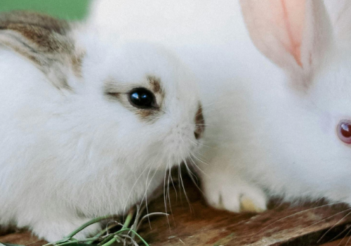
| Susceptibility | Rabbits are generally not a known host for this virus SARS-CoV-2. However, studies have found rabbits have mild to moderate ACE2 binding, and can be experimentally infected with high-dose inoculums. Rabbits have not been a significant part of SARS-2 research to date. | Not reported for SARS-CoV-1 (rabbits were not tested during the SARS outbreak). | Rabbits can be experimentally infected with MERS-CoV. In trials, inoculated rabbits did become infected (virus detected in tissues) and developed an immune response. | There’s no prominent literature on HCoV-229E infection in rabbits. It’s not a standard model (rabbits have their own seasonal coronaviruses, but not 229E). | Rabbits are not known to be susceptible to OC43. | NL63 infection of rabbits has not been described. | HKU1 infection of rabbits has not been described. |
| Transmission | Transmission between rabbits has been poorly studied. | N/A (no infection) | Limited: While rabbits shed MERS-CoV from the nose (up to 7 days post infection), they show no symptoms, so active transmission to other rabbits has not been observed in the lab (it wasn’t specifically tested; direct contact might result in transmission given nasal shedding). | N/A (not studied) | N/A (no infection) | N/A (not studied) | N/A (not studied) |
| Shedding | In rabbits infected with SARS-CoV-2, infectious virus has been recovered from the nasal cavity for up to 7 days post-infection. | N/A (no infection) | Infectious MERS-CoV has been recovered from rabbit nasal swabs through day 7, and viral RNA from the throat through day 3. No virus was detected in feces or urine. Thus, rabbits primarily shed virus via the respiratory route, and only briefly. | N/A (not studied) | N/A (no infection) | N/A (not studied) | N/A (not studied) |
| Symptoms and Pathology | Infected rabbits show no clinical signs of disease. Mild lung inflammation can be detected. | N/A (no infection) | Infected rabbits remain free of clinical signs (no fever or weight loss). Histopathology shows mild rhinitis and focal mild inflammation in nasal turbinates. There is minimal to no lung pathology (only low levels of virus in lungs). Rabbits do seroconvert by 21 days post-infection, confirming infection took place. Overall, rabbits experience an asymptomatic or very mild upper-respiratory infection with MERS-CoV. | N/A (not studied) | N/A (no infection) | N/A (not studied) | N/A (not studied) |
| Ethics and Logistics | Rabbits are an inefficient model for SARS-CoV-2. Due to the wide availability of small animal models, and the ethical restraints of using larger models, there is little justification for rabbit model use for SARS-CoV-2. | N/A (no model) | Rabbits are relatively easy to handle and are cheaper than NHPs. Because they do not get sick, they represent a subclinical carrier model for MERS. This is useful for testing vaccines (rabbits were used to see if they produce neutralizing antibodies) but limited for studying disease pathology or transmission (since they don’t develop nor spread a fulminant infection). BSL-3 is required. | N/A (no model) | N/A (no model) | N/A (no model) | N/A (no model) |
Rabbits: References
Baseler, L., E. de Wit, and H. Feldmann. 2016. “A Comparative Review of Animal Models of Middle East Respiratory Syndrome Coronavirus Infection.” Veterinary Pathology 53 (3): 521–31. https://doi.org/10.1177/0300985815620845.
Brand, J.M.A. van den, B.L. Haagmans, L.B. Provacia, V.S. Raj, K.J. Stittelaar, S. Getu, L. de Waal, et al. 2015. “Mers Coronavirus Infection of Rabbits.” Journal of Comparative Pathology 152 (1): 53. https://doi.org/10.1016/j.jcpa.2014.10.051.
Doremalen, Neeltje van, and Vincent J. Munster. 2015. “Animal Models of Middle East Respiratory Syndrome Coronavirus Infection.” Antiviral Research 122 (October):28–38. https://doi.org/10.1016/j.antiviral.2015.07.005.
Mykytyn, Anna Z., Mart M. Lamers, Nisreen M. A. Okba, Tim I. Breugem, Debby Schipper, Petra B. van den Doel, Peter van Run, et al. n.d. “Susceptibility of Rabbits to SARS-CoV-2.” Emerging Microbes & Infections 10 (1): 1–7. https://doi.org/10.1080/22221751.2020.1868951.
| Domestic cats | SARS-CoV-2 (COVID-19) | SARS-CoV-1 (SARS) | MERS-CoV (MERS) | HCoV-229E (Seasonal) | HCoV-OC43 (Seasonal) | HCoV-NL63 (Seasonal) | HCoV-HKU1 (Seasonal) |
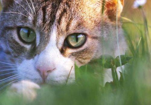
| Susceptibility | Domestic cats are permissive to SARS-CoV-2 infection but disease is typically mild. | Domestic cats are susceptible to SARS-CoV-1, but disease is mild or non-existent. | Cats have not been reported as naturally or experimentally infected with MERS-CoV. (Non-permissive DDP4). | Cats are not known to contract HCoV-229E. | Cats are not hosts for OC43. | Cats are not hosts for NL63. | Cats are not hosts for HKU1. |
| Transmission | High cat-cat transmission: The virus spreads via respiratory droplets when cats are in close contact (e.g. shared cages). There is no evidence, however, of cats transmitting to humans under normal conditions. | High cat-cat transmission, similar to SARS-CoV-2. | N/A (no infection) | N/A (no infection) | N/A (no infection) | N/A (no infection) | N/A (no infection) |
| Shedding | Domestic cats shed SARS-CoV-2 from the nasal and oral cavities. Viral RNA and infectious virus have been detected in feline oropharyngeal swabs and feces. Shedding in experimentally infected cats began ~2 days post-infection and lasted ~ up to 10 days. | Domestic cats shed SARS-CoV from the throat/nose starting ~2 days post-infection and continuing for about 10 days. Viral RNA was detected in nasal and pharyngeal swabs until day 10. (Cats also shed virus in feces in some cases, although respiratory droplet is the main route.) | N/A (no infection) | N/A (no infection) | N/A (no infection) | N/A (no infection) | N/A (no infection) |
| Symptoms and Pathology | Most domestic cats remain asymptomatic or develop very mild respiratory signs. (In studies, infected cats did not appear sick despite robust viral replication in the nasopharynx.) Pathology in cats is minimal; they do not develop the pneumonia seen in humans. | Infected cats showed no clinical signs despite active infection. Pathological examination of a few sacrificed cats at 4 days post-infection showed virus in respiratory tissues but relatively mild lesions. Thus, cats experience a subclinical infection while shedding virus. | N/A (no infection) | N/A (no infection) | N/A (no infection) | N/A (no infection) | N/A (no infection) |
| Ethics and Logistics | Cats are not standard lab animals, but their demonstrated susceptibility prompted concern in the COVID-19 pandemic. Experimentally, only small numbers are used and must be housed in containment. Ethical considerations are significant since cats are companion animals; studies are done with care to avoid suffering (which is generally minimal given mild disease). | Cats in SARS studies were euthanized for tissue analysis; as companion animals, their use raises ethical issues. Because they showed no illness, the model is one of asymptomatic carrier transmission, relevant to understanding potential silent spread. BSL-3 is required. | N/A (no infection) | N/A (no infection) | N/A (no infection) | N/A (no infection) | N/A (no infection) |
Domestic cats: References
Cleary, Simon J., Simon C. Pitchford, Richard T. Amison, Robert Carrington, C. Lorena Robaina Cabrera, Mélia Magnen, Mark R. Looney, Elaine Gray, and Clive P. Page. 2020. “Animal Models of Mechanisms of SARS‐CoV‐2 Infection and COVID‐19 Pathology.” British Journal of Pharmacology 177 (21): 4851–65. https://doi.org/10.1111/bph.15143.
Gaudreault, Natasha N., Jessie D. Trujillo, Mariano Carossino, David A. Meekins, Igor Morozov, Daniel W. Madden, Sabarish V. Indran, et al. n.d. “SARS-CoV-2 Infection, Disease and Transmission in Domestic Cats.” Emerging Microbes & Infections 9 (1): 2322–32. https://doi.org/10.1080/22221751.2020.1833687.
Shi, Jianzhong, Zhiyuan Wen, Gongxun Zhong, Huanliang Yang, Chong Wang, Baoying Huang, Renqiang Liu, et al. 2020. “Susceptibility of Ferrets, Cats, Dogs, and Other Domesticated Animals to SARS-Coronavirus 2.” Science (New York, N.Y.) 368 (6494): 1016–20. https://doi.org/10.1126/science.abb7015.
| Palm civets | SARS-CoV-2 (COVID-19) | SARS-CoV-1 (SARS) | MERS-CoV (MERS) | HCoV-229E (Seasonal) | HCoV-OC43 (Seasonal) | HCoV-NL63 (Seasonal) | HCoV-HKU1 (Seasonal) |
| Susceptibility | No evidence of SARS-CoV-2 infection in civets. | Masked palm civets, the implicated intermediate host in the 2003 SARS outbreak, are highly susceptible to SARS-CoV-1. In experimental infections, all civets became infected and developed high viral loads. | No evidence of MERS-CoV infection in civets. | No evidence of HCoV-229E infection in civets. | No evidence of HCoV-OC43 infection in civets. | No evidence of HCoV-NL63 infection in civets. | No evidence of HCoV-HKU1 infection in civets. |
| Transmission | N/A (not studied) | In nature, civets in live markets transmitted SARS-CoV to humans (e.g., in a restaurant outbreak). Although dedicated lab transmission studies aren’t detailed, infected civets shed virus copiously via respiratory routes, so droplet transmission between civets is probable. | N/A (not studied) | N/A (not studied) | N/A (not studied) | N/A (not studied) | N/A (not studied) |
| Shedding | N/A (not studied) | Infected civets shed SARS-CoV from respiratory and gastrointestinal tracts. Viral RNA has been found in civet lung, intestine, and brain tissues, indicating systemic spread; nasal/oral shedding would be expected during acute infection (as evidenced by transmission to handlers). | N/A (not studied) | N/A (not studied) | N/A (not studied) | N/A (not studied) | N/A (not studied) |
| Symptoms and Pathology | N/A (not studied) | Civets develop an illness resembling human SARS. They show fever (pyrexia), lethargy, and diarrhea. Pathology is multi-organ: civets get interstitial pneumonia, spleen and lymph node damage, hepatitis, and encephalitis, mirroring lesions seen in severe human cases. This similarity suggests civets suffer a SARS-like disease. | N/A (not studied) | N/A (not studied) | N/A (not studied) | N/A (not studied) | N/A (not studied) |
| Ethics and Logistics | N/A (not studied) | Civets are exotic animals requiring special permits and housing. They can be aggressive (they become less aggressive when sick). Using civets in research is challenging and raises ethical issues, but they provide a valuable pathogenesis model for SARS (insight into multi-organ effects). Today, civet use is rare, confined to understanding zoonotic transmission. BSL-3 is required. | N/A (not studied) | N/A (not studied) | N/A (not studied) | N/A (not studied) | N/A (not studied) |
Palm civets: References
Sutton, Troy C., and Kanta Subbarao. 2015. “Development of Animal Models against Emerging Coronaviruses: From SARS to MERS Coronavirus.” Virology 479 (May):247–58. https://doi.org/10.1016/j.virol.2015.02.030.
Wu, Donglai, Changchun Tu, Chaoan Xin, Hua Xuan, Qingwen Meng, Yonggang Liu, Yedong Yu, et al. 2005. “Civets Are Equally Susceptible to Experimental Infection by Two Different Severe Acute Respiratory Syndrome Coronavirus Isolates.” Journal of Virology 79 (4): 2620–25. https://doi.org/10.1128/JVI.79.4.2620-2625.2005.
| Dromedary camels | SARS-CoV-2 (COVID-19) | SARS-CoV-1 (SARS) | MERS-CoV (MERS) | HCoV-229E (Seasonal) | HCoV-OC43 (Seasonal) | HCoV-NL63 (Seasonal) | HCoV-HKU1 (Seasonal) |
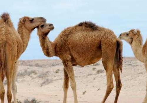
| Susceptibility | Camels are not known hosts for SARS-CoV-2. | Camels are not known hosts for SARS-CoV-1. | Dromedary camels are the natural reservoir host for MERS-CoV. They are readily infected and often seropositive in endemic regions. Experimental inoculation of camels with MERS-CoV leads to reliable infection. | Suspected origin host. HCoV-229E is believed to have ancestral links to camels or camelids (a related virus infects camels and alpacas), but modern HCoV-229E primarily circulates in humans. Camels can be infected by 229E-like viruses in nature, though experimental infection data is lacking. | Camels are not known hosts for OC43 (OC43 likely jumped from cattle in the 19th century). | Camels are not known to be susceptible. | Camels are not known to be susceptible. |
| Transmission | N/A (not studied) | N/A (not studied) | Camels transmit MERS-CoV via respiratory droplets. In barn settings, infected camels have passed the virus to other camels and to humans in close contact. Camels exhale viral particles (viral RNA detected in exhaled breath) and nasal discharge, enabling droplet spread. | Camels are not used in 229E lab studies, but if they were a source, transmission to humans would presumably have been by respiratory routes. | N/A (not studied) | N/A (not studied) | N/A (not studied) |
| Shedding | N/A (not studied) | N/A (not studied) | Camels readily shed MERS-CoV from their upper airways. Infectious virus is recoverable from nasal swabs for ~7 days post-infection, and viral RNA can persist in the nose for up to 35 days. Oropharyngeal shedding is lower and shorter duration than nasal shedding. | N/A (not studied) | N/A (not studied) | N/A (not studied) | N/A (not studied) |
| Symptoms and Pathology | N/A (not studied) | N/A (not studied) | Infected camels exhibit mild upper respiratory illness with occasional transient fever. The infection is largely restricted to the upper respiratory tract: viral antigen is found in nasal turbinate epithelium and trachea, with much less in lung tissue. Camels generally recover uneventfully and develop neutralizing antibodies by about 2 weeks post-infection. | N/A (not studied) | N/A (not studied) | N/A (not studied) | N/A (not studied) |
| Ethics and Logistics | N/A (no model). | N/A (no model). | Camels are large livestock, so experiments are costly and require specialized, large BSL-3 facilities. Ethically, disease in camels is mild, but their use is limited to select studies. Despite challenges, camels are an authentic transmission model for MERS (reflecting how the virus spreads at the animal source). | N/A (not studied) | N/A (not studied) | N/A (not studied) | N/A (not studied) |
Dromedary camels: References
Azhar, Esam I., Sherif A. El-Kafrawy, Suha A. Farraj, Ahmed M. Hassan, Muneera S. Al-Saeed, Anwar M. Hashem, and Tariq A. Madani. 2014. “Evidence for Camel-to-Human Transmission of MERS Coronavirus.” The New England Journal of Medicine 370 (26): 2499–2505. https://doi.org/10.1056/NEJMoa1401505.
Doremalen, Neeltje van, and Vincent J. Munster. 2015. “Animal Models of Middle East Respiratory Syndrome Coronavirus Infection.” Antiviral Research 122 (October):28–38. https://doi.org/10.1016/j.antiviral.2015.07.005.
